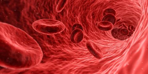
“With the launch of the new Bacteriophage Journal in early 2011, Alexander Sulakvelidze defined bacteriophages as “the most ubiquitous organisms on Earth, playing a vital role in maintaining the microbial balance on this planet. Indeed, bacteriophages, or phages, are found wherever their bacterial host is present. It has been determined that the phage population in water systems ranges from 10^4 to 10^8 virions per ml and approximately 10^9 virions per g in soil.” . FEMS Microbiol with an estimated total of 10^32 bacteriophages on the planet.
Bacteriophages: an assessment of their role in the treatment of bacterial infections. Phages, described almost a century ago by William Twort and independently discovered shortly thereafter by Félix d’Herelle (considered by many to be the founder of bacteriophages and their therapeutic significance: phage therapy), are small viruses that have the ability to kill bacteria without affecting the cell lines of other organisms. Due to the specificity of cellular target hosts, the application of phages has been proposed as a therapy for treating acute and chronic infections since their introduction, with initial successes first described in the disciplines of dermatology, ophthalmology, urology, stomatology, pediatrics, ENT, and surgery.
The initial enthusiasm for phage therapy to treat bacterial diseases in the pre-antibiotic era was understandably enormous. Indeed, the only therapy available in the 1920s and 1930s was serum therapy for selected pathogens such as pneumococci and diphtheria. The use of bacteriophages was even described with great interest when the protagonist of Sinclair Lewis’s Pulitzer Prize-winning novel, Arrowsmith, used this treatment to combat the outbreak of bubonic plague on a Caribbean island.
However, this concept of the therapeutic use of phages for treating bacterial infections was highly controversial from the outset and not widely accepted by the public and the medical community. Early studies were often criticized for lacking appropriate controls and inconsistent results. The lack of reproducibility and the many conflicting results obtained in various published studies led to the conclusion that the evidence for the therapeutic value of lytic filtrates was largely contradictory and inconclusive, and further research was recommended to confirm its alleged benefits. The emergence of the antibiotic chemotherapy era with the introduction of sulfa drugs in the 1930s and later penicillin in the 1940s further dampened enthusiasm for phage research and therapy. Notably, over the past decade, due to the emergence of multidrug-resistant bacteria, researchers have revisited this century-old approach, considering phage therapy as a “new” and potentially viable treatment option for hard-to-treat bacterial pathogens.
This essay will discuss the origins of phage therapy, the biology and life cycle of phages, and a summary of experimental and clinical data supporting phage therapy in the treatment of multidrug-resistant (MDR) bacterial infections and sepsis. It remains to be seen whether phage therapy will ever reach its full therapeutic potential in modern intensive care, but its practical applicability as an alternative to antibiotics for treating human sepsis caused by pathogens carrying multiple antibiotic resistance genes is now being seriously considered.
Historical Background
In 1896, Ernest Hanbury Hankin, a British bacteriologist serving as chemical examiner and bacteriologist for the Government of the United Provinces and Central Provinces of India, demonstrated that the waters of the Indian rivers Ganga and Yamuna contained a biological principle that destroyed cultures of cholera-causing bacteria. This substance could pass through Millipore filters, which are known to retain larger microorganisms like bacteria. He published his work in French in the Annales de l’Institut Pasteur. While studying the growth of vaccinia virus on cell-free agar media in 1915, British microbiologist Frederick Twort observed that “pure” bacterial cultures could be associated with a permeable filterable material, which could potentially cause bacteria from a culture to be completely disintegrated into granules. This “filterable agent” was detected in cultures of micrococci isolated from vaccinia: material from some colonies that could not be subcultured was able to infect new growth of micrococci, and this condition could be transferred to fresh cultures of the microorganism for a short, almost indefinite number of generations. This transparent material, which was found unable to grow without bacteria, was described by Twort as a ferment excreted by the microorganism for a purpose not clear at the time.
Two years after this report, Félix d’Herelle independently described a similar experimental finding while studying patients suffering from or recovering from bacillary dysentery. He isolated a so-called “anti-Shiga microbe” from the stools of shigellosis patients by filtering stools incubated for 18 hours. This active filtrate, when added to either a culture or an emulsion of Shiga bacilli, could cause the cessation of culture growth, death, and eventually lysis of the bacilli. D’Herelle described his discovery as a microbe that was a “true” microbe of immunity and an obligate bacteriophage. He also demonstrated the activity of this anti-Shiga microbe by inoculating laboratory animals for the treatment of shigellosis and appeared to confirm the clinical significance of his finding by satisfying at least some of Koch’s postulates.
Aside from the actual discussion about d’Herelle’s origin (some state he was born in Paris, while others claim Montreal), the initial controversy was mainly led by Bordet and his colleague Gartia at the Pasteur Institute in Brussels. These authors made competing claims about the exact nature and significance of the fundamental discovery. While Twort, due to lack of funds and his affiliation with the Royal Army Medical Corps, did not continue his research in the same field, d’Herelle introduced the use of bacteriophages in clinical medicine and published many non-randomized studies from around the world. He also conducted treatment with intravenous phages for invasive infections, and he summarized all these findings and observations in 1931. However, the first published article on the clinical use of phages was in Belgium by Bruynoghe and Maisin, who used bacteriophages to treat cutaneous furuncles and carbuncles by injecting Staphylococcus-specific phages near the base of the skin boils. They described clear evidence of clinical improvement within 48 hours with a reduction in pain, swelling, and fever in the treated patients.
At that time, the exact nature of the phage was not yet known, and it remained a matter of active and lively debate. The lack of knowledge about the essential nature of DNA and RNA as the genetic essence of life prevented a more comprehensive understanding of phage biology in the early 20th century. In 1938, John Northrop concluded from his own work that bacteriophages were produced by living hosts through the generation of an inert protein, which is converted into the active phage by an autocatalytic reaction.
However, several contributions from other researchers supported d’Herelle’s idea that phages were living particles or viruses when they replicated within their host cells. In 1928, Wollman assimilated the properties of phages with those of genes. In 1925, Bordet and Bail confirmed the idea that the ability of phages to reproduce in bacteria necessitates the insertion of phage-encoded material into the hereditary units of the host microbe. Frank Macfarlane, an Australian scientist who received the Nobel Prize in 1960 for his work on immunity, also worked on lysogeny and confirmed the viral nature of phages as well as the nature of their interactions with bacterial hosts. He also demonstrated that different types of phages exist.
Finally, the invention of the electron microscope (EM) allowed German physician Helmut Ruska to first describe spherical particles as well as “sperm-shaped” particles from a phage suspension adhering to a bacterial membrane. Two years later, in his dissertation, he summarized his most important research on the nature and biology of bacteriophages. A year after the first description of phages with EM, Luria and Anderson in Camden, New Jersey, presented various types of phages and described their common structure: an inhomogeneous spherical head with a much thinner tail, which gives the peculiar sperm-like appearance. They also described the different stages of bacterial lysis: increasing adsorption over time, extensive bacterial damage, and the appearance of a large number of newly formed bacteriophages.
While phage research was never abandoned in the former USSR with the development of the Eliava Institute in Tbilisi, Georgia, and some other countries like Poland (and the well-known Hirszfeld Institute in Wroclaw), English literature rediscovered phage therapy in animals in the 1980s, and human trials began in the 2000s, with the first Phase I randomized study published in the USA in 2009.
In August 2004, the so-called Phage Summit took place in Key Biscayne, Florida. Over 350 conference participants attended this first major international meeting in decades dedicated to phage biology. Overall, phage literature has become one of the most extensive topics, making bacteriophages one of the most thoroughly studied microbes known to science. In 1958 and 1967, Raettig published two bibliographies with approximately 11,358 references. In 2012, Ackerman analyzed 30,000 phage publications published between 1965 and 2010. The names of the first authors represent 40 language domains or geographical areas and at least 70 languages, leading to the conclusion that phage particles are studied worldwide (even if English and German languages predominate).
Types of Phages and Phage Biology
More than 6,000 different bacteriophages have been discovered and morphologically described, including 6,196 bacterial and 88 archaeal viruses. The vast majority of these viruses are attenuated, while a small portion are polyhedral, filamentous, or pleomorphic. They can be classified by their morphology, genetic content (DNA vs. RNA), specific host (for example, the Staphylococcus phage family, the Pseudomonas phage family, etc.), the location where they live (marine virus compared to other habitats), and their life cycle (see below). Over time, new classification formats have been proposed, and abbreviations for these viruses were suggested by Fauquet and Pringle in 2000.
As an obligate intracellular parasite of a bacterial cell, phages exhibit various life cycles within the bacterial host: lytic, lysogenic, pseudolysogenic, and chronic infection.
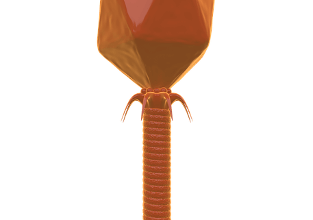
In phage therapy, the main interest has been in lytic phages, primarily represented in three families of the Caudovirales order: the Myoviridae, the Siphoviridae, and the Podoviridae. There are also some reports of applications of cubic phages and filamentous phages. The general description of these phages can be summarized as follows: The genetic material is contained within a protein shell or capsid, which has the shape of an icosahedron; this head is connected via a collar to the tail, which may or may not be contractile, and whose distal end is in contact with tail fibers whose tips recognize attachment sites on receptors of the bacterial cell surface.
Regardless of the type of phage life cycle, the first step is binding to receptors on the bacterial cell wall before phages can penetrate the bacteria. This specific process influences the spectrum of possible interactions between phages and bacteria. For example, bacteriophage λ only interacts with the LamB receptor of E. coli. Spatiotemporal dynamics have shown that this event is of great importance for successful bacterial invasion. Some phages are also capable of synthesizing specific enzymes (such as hydrolases or polysaccharidases and polysaccharide lyases) that can break down exopolysaccharide structure capsules before they can interact with their specific
receptor. This is the case for some phages that interact with strains of E. coli, V. cholerae, P. aeruginosa, E. agglomerans, and P. putida. These enzymes are of potential interest for their therapeutic implications and are currently in preclinical development.
Upon binding to its specific receptor, phages induce a pore in the bacterial cell wall and inject their DNA into the cell, while the viral capsid remains outside the bacteria. This is followed by the expression of early phage genes, which, in the case of lytic phages, redirect the bacterial synthetic machinery towards the reproduction of viral nucleic acids and proteins. The assembly and packaging of phages are then observed before the lysis of bacterial cells and the release of phage progeny occur. The late enzymes of phages, such as lysins, holins, and inhibitors of murein synthesis, are then used for the virion burst into the extracellular environment. The number of released viral particles or the size of the bursts varies greatly depending on the phage, the state of the bacterial host, and other environmental factors, such as nutrient components surrounding the host.
In the lysogenic cycle, so-called temperate phages insert their genetic content (the prophage) into the bacterial chromosomes, where they remain dormant for extended periods and are replicated as part of the bacterial chromosome. Therefore, there is no self-replication. This prophage DNA is vertically transmitted along with the entire bacterial genome to its progeny until the lytic cycle is induced.
During induction, lysogenic phage can occasionally transfer host genetic material adjacent to its insertion site on the chromosome from one bacterium to another, a phenomenon known as transduction. In fact, the significant role of phages in the evolution of the bacterial genome has been known for years, and Brussow even described bacteriophages as a means for lateral gene transfer.
This process can promote the transfer of genes that are of selective advantage to the bacterial host, including antibiotic resistance genes; however, the same process could be therapeutically exploited by using phages to transfer genes that make bacteria more susceptible to some antibiotics. Indeed, Lu and Collins showed in vitro an increased sensitivity of E. coli to antibiotics by targeting DNA repair mechanisms through the injection of a specific gene that led to the overexpression of a protein inhibiting this system. Gene insertion was achieved by a specific and modified bacteriophage M13. Interestingly, they also used the same technique in mice infected intraperitoneally with E. coli. Survival was increased in mice treated simultaneously with antibiotics and modified phages. This approach was found by other authors to be similar to the general approach of phage therapy, which leads to the direct killing of bacteria.
Another approach is to reverse resistance to pathogens by injecting specific genes for a sensitization cassette that dominantly confers susceptibility. This was recently shown by Edgar and colleagues, who were able to make resistant bacteria susceptible to streptomycin and nalidixic acid.
Finally, chronic infection occurs when the bacterium is infected by lysogenic phages that subsequently mutate and lose the ability to trigger a lytic replication cycle. The phage DNA becomes a new part of the bacterial chromosome and becomes a long-term prophage sequence.
Why Do We Need Phage Therapy?
Over the past two or three decades, the widespread emergence and spread of antibiotic-resistant bacteria worldwide has become a major therapeutic challenge.
For example, MRSA infections in the USA were reported with an incidence of approximately 100,000 severe infections in 2005, leading to 20,000 deaths.
The limited therapeutic options for treating the most important multidrug-resistant bacteria (MDR), known by the acronym ESKAPE pathogens (for Enterococcus faecium, Staphylococcus aureus, Klebsiella pneumoniae, Acinetobacter baumannii, Pseudomonas aeruginosa, and Enterobacter spp.), have now become a looming health crisis in many intensive care units worldwide.
The treatment of patients with MDR pathogens has been shown by Morales et al. to increase the overall cost of care and prolong hospital stays.
In all healthcare professions, there is an ethical imperative to do everything in our power to preserve the effectiveness of antibiotics and to recognize that this precious resource is being wasted through the often unnecessary and inappropriate use of antibiotics, thereby promoting the acquisition and spread of antibiotic resistance genes. Antibiotic resistance is now considered a healthcare emergency, and many are calling for the development of new means to combat it. However, antibiotics are not developed based on direct public benefit, but on free-market criteria. Despite calls for the development of new antibiotics in the European Union (EU) and the United States (USA),
the World Medical Association’s statement on antimicrobial resistance (www.wma.net/e/policy/a19htm) indicates a lack of new antibiotics in the development pipeline.
A completely new, non-antibiotic approach to treating bacterial pathogens is certainly needed. The re-application of phage therapy could be a welcome alternative to antimicrobial chemotherapy in this phase of progressive spread of bacterial MDR pathogens with a lack of new antibiotics to combat these pathogens.
Furthermore, the need for phage applications certainly exceeds their use in human infections. Indeed, the use of bacteriophages has been described in various situations, including (but not limited to): food safety,
veterinary applications, and clinical diagnostic applications such as detection and typing of bacteria in human infections.
Potential Benefits of Phage Therapy
Bacteriophages are natural antibiotics capable of regulating bacterial populations by inducing bacterial lysis. They are active against gram-positive as well as gram-negative bacteria.
Since the mechanism of action of phage lysis is completely different from that of antibiotics, activity against bacteria that exhibit multiple mechanisms of antibiotic resistance is maintained.
Due to their specificity, phage therapy has a narrow antibacterial spectrum with an effect limited to a single species or, in some cases, a single strain within a species. This limits the “pressure” and severe collateral damage that occurs to bystander, non-targeted bacteria from antibiotics. The patient’s entire microbiome is altered by antibiotics, not just the intended target pathogen. Chibani-Chennoufi et al. showed only minor effects on the gut microbiota in mice after oral administration of E. coli-targeted phage therapy. The preservation of much of the existing microbiome during phage therapy has been confirmed in careful microbial surveys of adult healthy volunteers who received a 9-phage cocktail.
Phage therapy also avoids the potential overgrowth of secondary pathogens.
As there are currently no large, randomized, controlled trials, it is difficult to assess side effects and their potential impact. Based on reports from Poland and the former Soviet Union, phage therapy appears to be without significant adverse effects. The fact that bacteriophages only interact with bacterial cells and do not interfere with mammalian cells could potentially explain the lack of harmful side effects. Underreporting could be another explanation. However, the excellent tolerability of phage treatment has been demonstrated in preclinical studies in various animal models and in several observational studies in patients and healthy subjects. Systemic administration leads to a wide distribution of phages, including the ability to cross the blood-brain barrier, allowing these agents to be used in central nervous system infections.
Interestingly, at least some phages also show the ability to destroy bacterial biofilms.
Phage therapy can influence the inflammatory response to an infection. In 51 patients with various long-term suppurative infections, TNFα release in vivo and in vitro was attenuated after stimulation with LPS, based on the initial pattern of TNFα serum levels. IL-6 release was significantly reduced only in vivo. C-reactive protein and leukocyte count were initially unaffected in this patient population, while they significantly decreased between day 9 and day 32 in 37 patients receiving oral phage therapy for osteomyelitis, joint prosthesis infection, skin and soft tissue infections, and in one case, lung infection.
This was an observational study without a control group and should therefore be interpreted with caution. In a more recent observation, CRP was only affected in patients whose initial CRP serum level was above 10 mg/dL.
White blood cells can also be affected by phage therapy: in patients after 3 weeks and 3 months of therapy, increased neutrophil precursors and a decreased phagocytosis index for Staphylococcus aureus were observed compared to healthy donors. Recently, an extensive review of the alteration of immune responses during phage therapy was published.
Finally, the economic aspects of phage therapy are promising. Despite the fact that the duration of treatment was significantly prolonged, the costs of phage therapy were lower than with conventional antibiotic treatment, as shown in 6 patients with various staphylococcal infections, including methicillin-resistant Staphylococcus aureus.
Above all, the fact that bacteriophages could have improved efficacy compared to antibiotics is the greatest hope for the future. Smith and colleagues first demonstrated this finding in the early 1980s when they induced a lethal E. coli infection in mice with a highly virulent strain expressing a K1 polysaccharide capsule.
A single intramuscular dose of anti-K1 phages was as effective as multiple streptomycin injections and superior to multiple intramuscular doses of tetracycline, ampicillin, chloramphenicol, or trimethoprim in curing the animals. To our knowledge, this observation has never been confirmed in human infections.
Potential Limitations and Disadvantages of Phage Therapy
Despite all the advantages summarized above, we are far from calling phages a “miracle cure” for treating any type of infection. Indeed, the optimal dose, route of administration, frequency, and duration of treatment still need to be determined before widespread clinical trials are considered.
The main disadvantage of phage therapy is the need to quickly and accurately determine the exact etiological microorganism causing the infection. The exquisite specificity of phage therapy against specific pathogens is a great advantage, but also a burden. A clinical sample must be isolated and cultured using standard microbiology diagnostic procedures to identify the pathogen before a specific bacteriophage solution can be defined and later administered to the patient. Innovations in rapid bacterial diagnosis using genomic methods or mass spectrometry could provide a solution. However, in most clinical microbiology laboratories and resource-limited healthcare facilities, this is a time-consuming process.
This problem could potentially be solved by using ready-to-use phage cocktails. The selection of potent phages from an available collection after phage typing of the isolated bacteria defines the so-called compound phage cocktail treatment. Finally, if no active, existing phage preparation is available against a severe pathogen, it can be isolated directly from the environment before being prepared for application.
For example, during the recent outbreak of E. coli O104:H4 in Germany, active lytic phages were found in the collection of the Eliava Institute (Georgia) as well as in the wastewater of the Brussels Military Hospital in Belgium.
The choice of bacteriophage for therapy is limited to lytic phages.
Indeed, lysogenic phages induce delayed lysis, which prevents the use of these phages in an acute infection. Although standardized methods for generating phage cocktails exist, there are no clear official guidelines.
The stability of viruses with regard to their susceptibility to various external and physical factors has recently been reviewed and could be responsible for some difficulties in producing stable solutions.
Another concern of phage therapy is the potential ability of bacteriophages to transfer DNA from one bacterium to another. This transfer of genetic material, or transduction, could be responsible for the transfer of pathogenicity determinants and virulence factors, which could lead to the development of a new microbe or even more resistant bacteria.
Therefore, the use of phages that are unable to package additional host DNA, or phages that use host DNA to synthesize their own DNA, would be preferred. This technique has already been successfully used in phage therapy.
The genome of many phages has been deciphered, and every month there are reports of newly identified gene sequences. However, we are far from knowing the gene of every type of phage, and the function of many of these genes is still unknown. For example, the ORFan genes found in some phages have no similarity to any other gene in the gene database. The role of these genes in promoting harmful side effects still needs to be clarified.
At the end of their antibacterial action, lytic phages induce the lysis of bacteria and release various bacterial substances such as endotoxin (LPS) from gram-negative bacteria. This can be responsible for various side effects on the host, such as the development of an inflammatory cascade leading to multiple organ failure. However, this potential problem also affects currently available rapidly bactericidal antibiotics.
Since they are viruses, bacteriophages can be regarded by the patient’s immune system as potential invaders and therefore rapidly eliminated from the systemic circulation by the reticuloendothelial system before they can accumulate in the spleen or liver, or they can be inactivated by adaptive immune defense mechanisms. This can lead to reduced efficacy with prolonged or repeated use.
Finally, the development of resistance mechanisms by the bacterial host, induced either by mutation and selection or by the acquisition of temperate phages, could lead to reduced efficacy of phages. There are at least 4 mechanisms that can be involved in bacterial resistance to a specific phage. Loss or lack of receptor, structural modification and/or masking of the receptor prevent phage adsorption to the bacteria and prevent the further ability to produce new phages. Receptor loss can occur when the cell surface composition is changed, as has been shown for Bordetella spp.
A structural modification was found for the E. coli protein TraT, which modifies the conformation of OmpA (Outer-Membrane Protein A), the receptor for T-Even-like phages. Secretion of various molecules (such as exopolysaccharide from Pseudomonas spp. or glycoconjugates from Enterobacteriaceae) can mask the receptor, but phages can counteract this by selecting a new receptor or by secreting the exopolysaccharide-degrading enzyme.
The other resistance mechanisms include preventing phage DNA integration by the Superinfection Exclusion System (Sie), degradation of phage DNA by the Restriction-Modification defense system or by Clustered Regularly Interspaced Short Palindromic Repeats (CRISPR), and blocking phage replication and transcription, translation, or virion assembly by the Abortive Infection system.
Fortunately, the frequency of resistance in vivo during phage therapy has so far been reported to be low, in contrast to the in vitro resistance analyses observed. Furthermore, the isolation of new active phages from the environment or the progressive isolation of “adapted” phages could offer a new treatment option.
In most countries, phage therapy is not covered by statutory health insurance, which represents a potential financial problem for some patients. There are some exceptions. The Swiss authorities have decided to reimburse the costs of complementary medicine for a period of 6 years while efficacy is being evaluated, and the President of the city of Wroclaw (where the Hirszfeld Institute is located), Poland, has created a program to cover the costs of phage therapy for the city’s residents; 2 examples to follow according to Myedzybrodzki.
Since bacterial viruses are currently not recognized as medicinal products, the current pharmacological regulations, definitions, and standards in Europe are not adequately adapted to phage preparations. For this reason, a Belgian research group and some members of the Pasteur Institute in Paris developed PHAGE (for Phages for Human Application Group Europe), an international non-profit organization, with the aim of developing a specific framework for the use of bacteriophages.
Regulatory approval remains another hurdle. In addition to safety concerns, neither the US Food and Drug Administration (FDA) nor the European Medicines Agency has an approval process that can easily accommodate the constantly changing phage combinations that companies need to develop to stay ahead of the development of MDR bacteria.
Experimental Data with Phage Therapy
Much experimental data has been generated since the two seminal studies by Smith and Huggins, which demonstrated the potential role of bacteriophages in combating systemic infections and enteritis in mice, calves, piglets, and lambs in the early 1980s.
Mice have been extensively studied as experimental animals, but there are also reports of phage therapies in laboratory models of infections in rats, chickens, rabbits, calves, and lambs. Various models of infections have been evaluated, such as intraperitoneal injection of live bacteria leading to systemic infection with bacteremia, intramuscular injection of bacteria, central nervous system infection, lung infection, liver abscesses, enteritis, urinary tract infection, bone infection, skin and wound infections. Bacteria used in these models included E. coli, MDR bacteria (Pseudomonas aeruginosa, ESBL-producing E. coli and K. pneumoniae, vancomycin-resistant Enterococcus faecium), Staphylococcus aureus, and Chronobacter turicensis. Some strains were isolated directly from patients. The tested administration procedures for phage therapy include intraperitoneal injection, oral or intragastric administration, topical, subcutaneous, and intramuscular injections, and intranasal administration. While in some studies phage administration was considered a prophylactic measure, treatment was usually administered as a single dose after bacterial challenge and in some studies was delayed until the animals showed symptoms of diarrheal infection or clear signs of severe infection.
Overall, these studies showed positive effects on mortality under phage therapy, and in 3 studies where mortality was assessed, the results were significantly better than with antibiotics used as comparative agents.
In one study of the infected bone model in rats, combined antibiotic-bacteriophage treatment significantly reduced the quantitative culture from the infected site at the end of the study compared to either treatment modality given alone.
Human Applications Already Described
In the first report on the use of bacteriophages in humans, efficacy was demonstrated in staphylococcal skin furuncles, and d’Herelle summarized all his clinical work in 1931. In the 1930s, there was a large number of publications and a complete monograph of the journal La Médicine dealt with phage applications in human diseases. The treatment of typhoid, Shigella, and Salmonella spp.-related colitis, peritonitis, skin infections, surgical infections (mainly abscesses of various locations), septicemia, urinary tract infections, and otolaryngological infections (external otitis and nasal furuncles) was described.
As previously described, enthusiasm for phage therapy in Western countries declined in the 1930s due to the reports by Eaton and colleagues and also as a consequence of the discovery and ease of use of antibiotics. The use of bacteriophages continued in Eastern countries, and over time numerous reports were published, primarily in Poland and Georgia (former USSR). The use of non-English literature (mainly Russian and Polish) likely explains the fact that these reports were limited to the authors’ countries of origin. A summary of this literature was recently published by various authors. However, we must note that most published data come from non-randomized, uncontrolled studies.
In fact, the first Phase I controlled randomized study conducted in the United States was published in 2009. It evaluated the safety of a cocktail of phages directed against E. coli, S. aureus, and Pseudomonas aeruginosa in 42 patients with chronic venous leg ulcers. The study could not demonstrate positive outcomes such as healing rate or frequency; however, the authors noted no adverse effects related to the treatment. Another randomized study was conducted in the United Kingdom and examined the efficacy of applying a solution containing 6 bacteriophages in the ears of patients with chronic Pseudomonas aeruginosa-related otitis. The number of P. aeruginosa colonies in the treated group in this well-conducted, double-blind, placebo-controlled study decreased significantly, while various subjective clinical indicators improved in these patients. In fact, patients reported reduced intensity of symptoms such as discomfort, itching, wetness, and unpleasant odor. Likewise, the physicians responsible for the patients (and blinded to the assigned treatment) reported reduced clinical observations such as erythema/inflammation, ulceration/granulation/polyps, and odors. No adverse effects were reported.
Recently, a small Phase I study was conducted with 9 patients treated at the Burn Wound Center of Queen Astrid Military Hospital in Brussels, Belgium. The patients were treated locally with the BFC-1 phage cocktail, which contained 3 lytic phages: a myovirus, a podovirus directed against Pseudomonas aeruginosa, and a myovirus directed against Staphylococcus aureus. A large burned section was exposed to a single spray application, while a distant part of the wound served as a control. While the complete results have yet to be published, no safety issues were reported.
Finally, a randomized controlled study confirmed the safety of an orally administered phage solution in healthy, non-infected patients.
Conclusions
Bacteriophages are a potential alternative tool for treating bacterial infections, including those caused by MDR pathogens. In fact, phage therapy has several advantages, and only few adverse events are reported, although underreporting cannot be excluded. However, further well-conducted studies are required to define the role and safety of phage therapy in daily clinical practice for treating patients with various infections.
Furthermore, the direct use of phage-encoded proteins such as endolysins, exopolysaccharidases, and holins has proven to be a promising alternative to antibacterial products. However, this topic would exceed the scope of this review.
Machine translation of the source: https://doi.org/10.4161/viru.25991
Xavier Wittebole, Sophie De Roock & Steven M Opal